Cross-section Microscopy Analysis: Mayo House Staircase Baluster Fragment and Staircase in situ at George Pitt House(Block 2, Building 28), Williamsburg, Virginia
Colonial Williamsburg Foundation Library
Research Report Series - 1744
Colonial Williamsburg Foundation
Library
Williamsburg, Virginia
2013
CROSS-SECTION MICROSCOPY ANALYSIS REPORT
Mayo House Staircase Baluster Fragment and Staircase in-situ at George Pitt House
COLONIAL WILLIAMSBURG FOUNDATION
WILLIAMSBURG, VIRGINIA
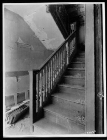 Figure 1. Mayo House stair in-situ, c.1929, before house demolition (CWF archives)
| Object: | Mayo House Staircase (Relocated Mayo House stair in George Pitt House and Mayo House stair baluster fragment in CWF collection [AF-CHES 90.0226]) |
|---|---|
| Requested by: | Edward Chappell, Roberts Director of Architectural and Archaeological Research; Colonial Williamsburg Foundation |
| Analyzed by: | Kirsten Travers, Graduate Fellow, Winterthur/University of Delaware Program in Art Conservation |
| Consulted: | Susan L. Buck, Ph.D., Conservator and Paint Analyst; Williamsburg, Virginia |
| Date submitted: | June 2011 |
Purpose:
The goal of this project is to use cross-section microscopy techniques to explore the finish history of the Mayo House staircase (fig. 1). Although the Mayo House was demolished in the early 20th-century, its original staircase was salvaged and installed in the reconstructed George Pitt House in Colonial Williamsburg (called the Pitt-Dixon House until the 1980s). Eight samples were collected from a Mayo House baluster fragment in the architectural fragments collection, and three samples were taken from the stair in-situ at the Pitt-Dixon house. It was also hoped that the comparative paint evidence would determine if any original finishes remained on the Pitt-Dixon stair, or if the stair was completely stripped of its earlier finishes during the installation.
History:
The George Pitt House, where the original Mayo House staircase is currently installed, is a reconstructed colonial dwelling located at the east end of Duke of Gloucester street (fig. 2). The original Pitt-Dixon house was constructed c.1718 and later occupied by notable Williamsburg personages such as George Pitt, an apothecary and surgeon who lived in the house from c 1754 - 1774, and John Dixon, a partner in the printing firm of Hunter and Dixon, located adjacent to the property. Dixon purchased the house from Pitt in 1774, but sold it six months later (Kocher and Dearstyne 1954, 11). A fire destroyed the house in the late 19th century. The present reconstruction was carried out by Colonial Williamsburg in 1936 based on archival and archaeological research, as well as historic photographs of the house as it stood in the late 19th century.
The main stair presently in the George Pitt house entrance hall originates from the Mayo House, a colonial dwelling which stood formerly on York street, block 8, near the Capitol (fig. 7). The building (for unknown reasons), was demolished during the reconstruction, but the stair was salvaged and re-installed in the reconstructed George Pitt House. The architectural report notes that the posts, rails, balusters and 3 even risers and treads were re-used (Kocher and Dearstyne 1954, 29). At that time a baluster was removed and added to the architectural fragments collection at CW (accession number AF-CHES 90.0226), which became the subject of the present analysis.
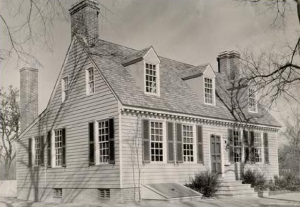 Figure 2. George Pitt House, reconstructed c.1936, southwest view, (CWF archives)
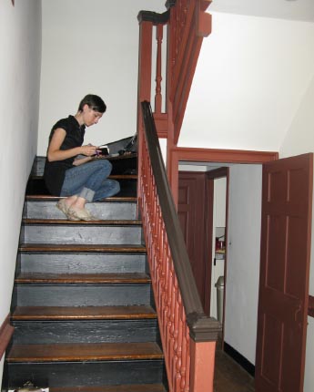 Figure 3. The author collects samples from the George Pitt House staircase (photo Dani Jaworski)
This fragment was originally the 14th baluster up from the first floor to the landing (see fig. 4). It is 27" tall and consists of a square base, a turned shaft, and a square cap (fig. 5). A significant accumulation of paints were observed on the fragment, which are extensively cracked and flaking. The present layer appears to be a very soiled white or cream-colored paint (fig. 6). A deep turquoise paint was visible beneath.
The paint history of this singular fragment may not necessarily reflect the overall finish history of the stair. An archival photograph of the interior of the Mayo House before demolition shows the staircase in-situ with light-colored balusters and a darker-colored newel post and handrail (fig. 8).
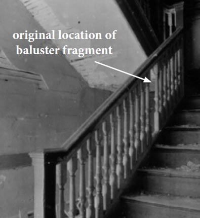 Figure 4. Detail of fig. 1, Mayo House stair before demolition. Note gap where baluster has been removed (CWF archives)
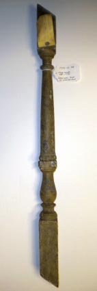 Figure 5. Mayo House baluster
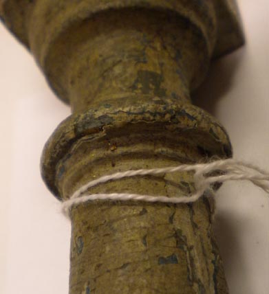 Figure 6. Detail of Mayo House baluster showing soiled and flaking painted surface
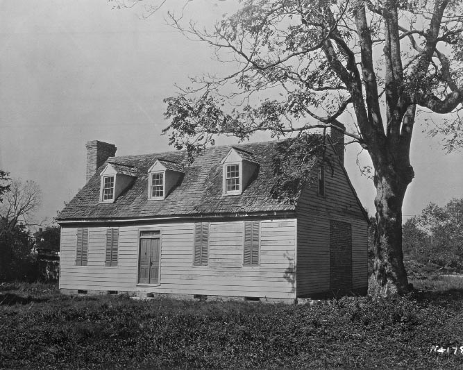 Figure 7. Mayo House exterior, before demolition, c.1929. (photo CWF archives).
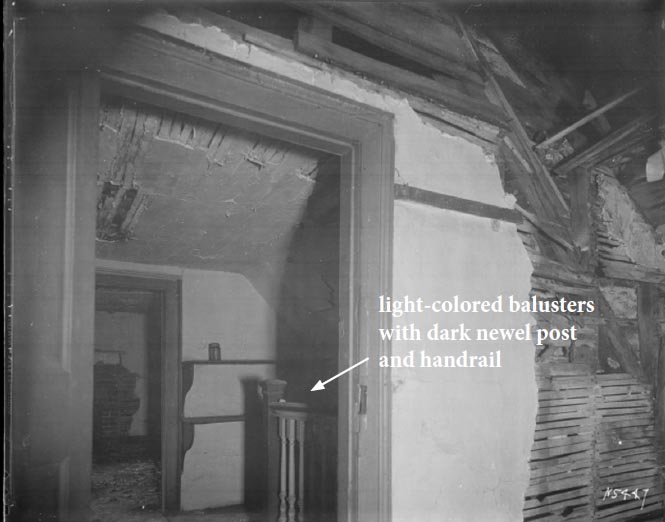 Figure 8. Mayo House interior, second floor, before demolition, c.1929. (photo CWF archives).
Procedures:
On September 27, 2010, the George Pitt House was visited by Kirsten Travers accompanied by Dani Jaworski (Architectural Collections Specialist) and Matthew Webster (Director of Historic Architectural Resources). The purpose of this visit was to collect samples from the stair to search for intact paint sequences that matched those found on the Mayo House baluster fragment. Three samples were collected (PHS 1-PHS 3). The George Pitt House stair is currently painted a glossy, deep red color with a dark brown handrail, and the current resident of the house related that the stair was previously painted a bright blue color (fig. 10). Small micro-excavations were made with a scalpel and examination at 30x with a monocular microscope found only these modern blue and red layers. Regardless, a microscalpel was used to remove three samples from the stair, and the location of sample sites was recorded and photographed. The samples were labeled and stored in small individual Ziploc bags for transport back to Susan Buck's paint analysis laboratory. All samples were given the prefix "PHS", and numbered according to the order in which they were collected.
On the same day, the baluster fragment from the Mayo House stair [AF-CHES 90.0226], was brought to the George Pitt House by Dani Jaworski and transferred to Kirsten Travers for examination and sampling. In the Architectural Research Department office, the baluster was examined, photographed, and sampled (fig. 9). Eight samples were taken and given the prefix "MHS" and numbered according to the order in which they were collected (MHS 1-MHS 8). A complete list of sample locations can be found in Table 1.
In the laboratory, the samples were examined with a stereomicroscope under low power magnification (5x to 50x), to identify those that contained the most paint evidence and would therefore be the best candidates for cross-section microscopy. Uncast sample portions were retained for future examination and analysis. The best candidates were cast in resin cubes and sanded and polished to expose the cross-section surface for microscopic examination. Please see Appendix A for sample preparation details.
| Sample number | Sample location | Sample description | Taken by/date |
|---|---|---|---|
| PHS 1 | George Pitt House stair in-situ | stair landing, half baluster against newel post, 'cap' of lower urnshape, currently deep red | Travers/September 27, 2010 |
| PHS 2 | George Pitt House stair in-situ | second flight, top molding of stringcourse, just below 6th baluster from landing, currently deep red | Travers/September 27, 2010 |
| PHS 3 | George Pitt House stair in-situ | second flight, top rail, fascia below cap, adjacent to newel post; currently dark brown | Travers/September 27, 2010 |
| MHS 1 | Mayo House baluster fragment [AF-CHES 90.0226] | top surface of base, 6 ½" up from bottom edge | Travers/September 27, 2010 |
| MHS 2 | Mayo House baluster fragment [AF-CHES 90.0226] | top surface of the 'urn' shaped element, 8" up from bottom edge | Travers/September 27, 2010 |
| MHS 3 | Mayo House baluster fragment [AF-CHES 90.0226] | top surface of shaft base, 12" up from bottom edge | Travers/September 27, 2010 |
| MHS 4 | Mayo House baluster fragment [AF-CHES 90.0226] | 'capital' cap of shaft, 21" up from bottom edge | Travers/September 27, 2010 |
| MHS 5 | Mayo House baluster fragment [AF-CHES 90.0226] | shaft, 19" up from bottom edge | Travers/September 27, 2010 |
| MHS 6 | Mayo House baluster fragment [AF-CHES 90.0226] | body of 'urn' shaped element, 8 ¾" up from bottom edge | Travers/September 27, 2010 |
| MHS 7 | Mayo House baluster fragment [AF-CHES 90.0226] | face of baluster base, 5 ¾" up from bottom edge | Travers/September 27, 2010 |
| MHS 8 | Mayo House baluster fragment [AF-CHES 90.0226] | face of baluster base, bottom edge | Travers/September 27, 2010 |
Sample location photographs (*indicates sample discussed in report)
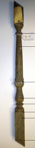 Figure 9. Mayo House stair Baluster fragment
[AF-CHES 90.0226]
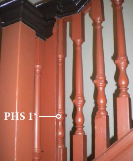 Figure 10. George Pitt
House stair samples
Figure 10a. Landing
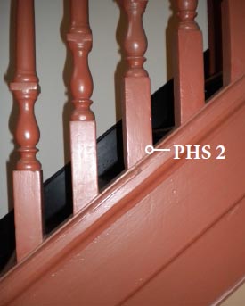 Figure 10b. Second flight, stringer
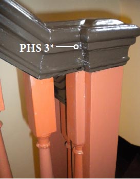 Figure 10c. Landing
Results:
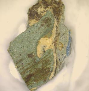
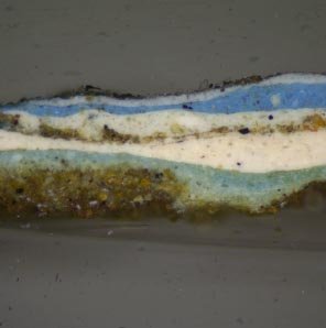
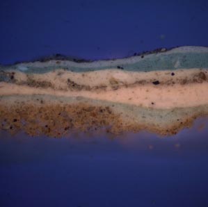
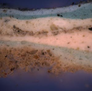
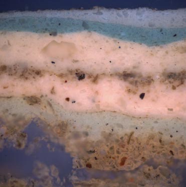
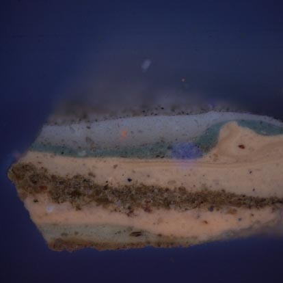
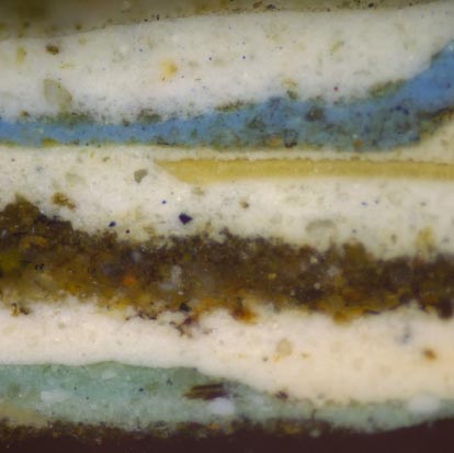
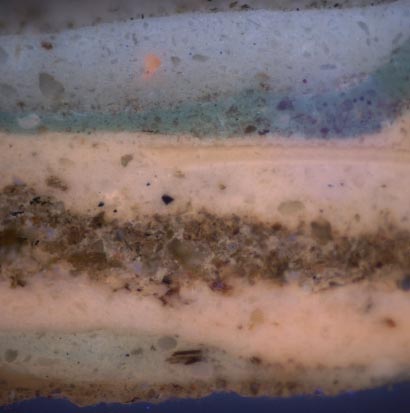
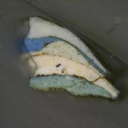
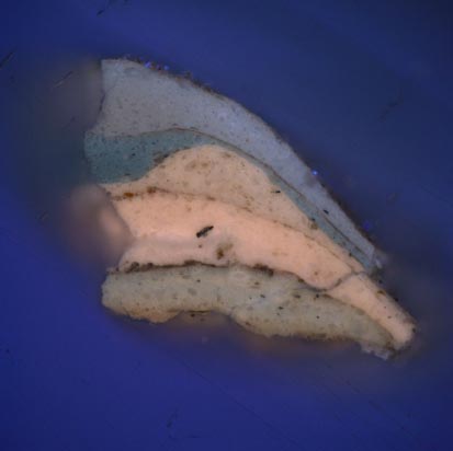
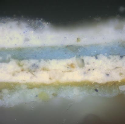
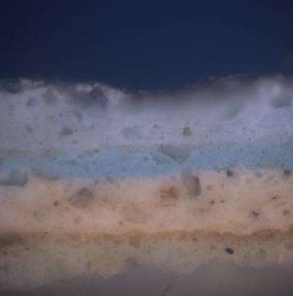
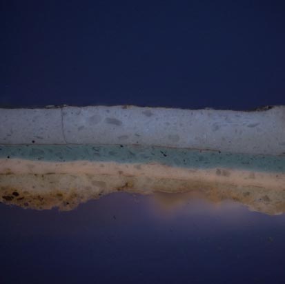
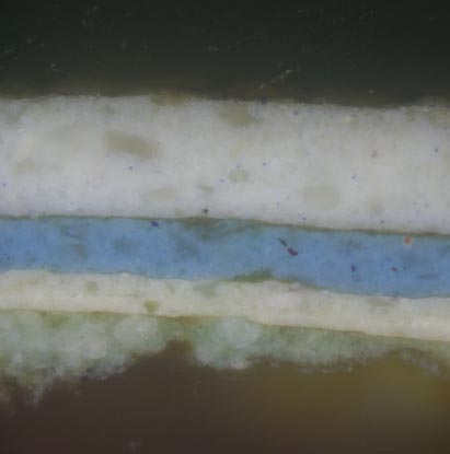
Seven finish generations were identified on the Mayo House baluster fragment, most of which consist of various shades of blue and white paints (table 2). The earliest coating appears to be a resinous layer, possibly shellac, which has a great deal of embedded particulate matter in its surface, suggesting it was exposed for a long period of time. Binding media analysis suggests this generation contains a carbohydrate component. The second generation is a greenish-blue paint that contains lead white, chalk, Prussian blue and yellow ochre pigments. Binding media analysis of this layer suggests it contains both oil and protein components. Generation 3 is a cream-colored paint, as is generation 4, which has a great deal of dirt accumulated on its surface. Generation five is a light blue paint, generation six is a deep blue paint, and generation seven (the current presentation surface), is a white paint. The same seven paint generations were found on all samples removed from the baluster fragment. To avoid redundancy, only cross-section samples MHS 1, MHS 2, and MHS 4 are discussed in the following section.
None of the pre-restoration paints on the baluster fragment were found on the stair in-situ at the George Pitt House, although a pale orange autofluorescence in the wood cells suggests that the early shellac coating may have survived. This was followed by a fragmented cream or gray-colored coating that may represent a paint that was not seen on the baluster fragment. The remaining paints currently on the Pitt- Dixon baluster and handrail all appeared to be modern, industrially prepared coatings, suggesting that the staircase was stripped of its finish history during the restoration.
The baluster fragment findings are discussed first. This section includes cross-section photomicrographs, binding media analysis with fluorochrome stains, and pigment identification with polarized light microscopy techniques. This is followed by a discussion of two samples collected from the staircase currently installed in the Pitt-Dixon house.
Where cross-section photomicrographs are shown, paint stratigraphies have been annotated according to finish "generation." For instance, a primer, paint layer, and varnish may represent one finish generation and are all given the same number, but differentiated with lowercase letters (1a, 1b, 1c, etc.), according to the order in which they would have been applied. Color matching is discussed in a separate section at the end of the report. All results are interpreted in the conclusion, and all sampling memoranda and raw photomicrographs can be found in the appendix at the back of this report.
| Generation | Description | Stain reactions | Pigments (PLM) | Notes |
|---|---|---|---|---|
| 7 | white paint | FITC+, DCF+ | n/a | current presentation surface, heavy soiling, discoloration, and flaking |
| 6 | deep blue paint | FITC+, DCF+ | n/a | — |
| 5 | light blue paint | FITC+, DCF+ | n/a | appears white in cross-section |
| 4 | cream paint | FITC+, DCF+ | n/a | heavily soiled |
| 3 | cream paint | FITC+, DCF+ | n/a | — |
| 2 | greenish-blue paint | faint FITC+, DCF+ | white lead, chalk, Prussian blue, yellow ochre | grime on surface |
| 1 | possibly resin coating with embedded dirt, orange autofl. suggests shellac | TTC+, possible FITC+ | n/a | solubilized in delivery solvents for fluorochrome stains |
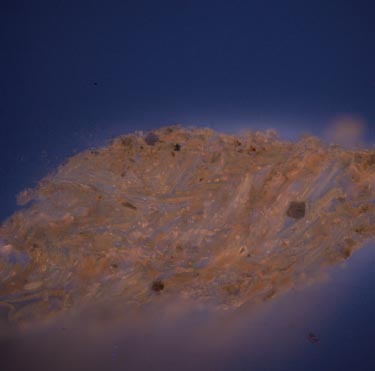
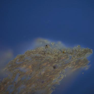
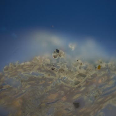
Sample MHS 5: Mayo House stair fragment
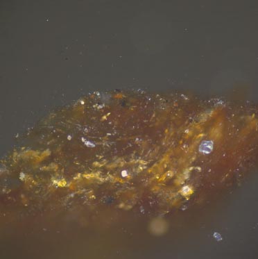
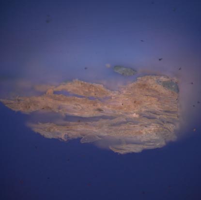
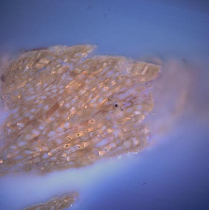
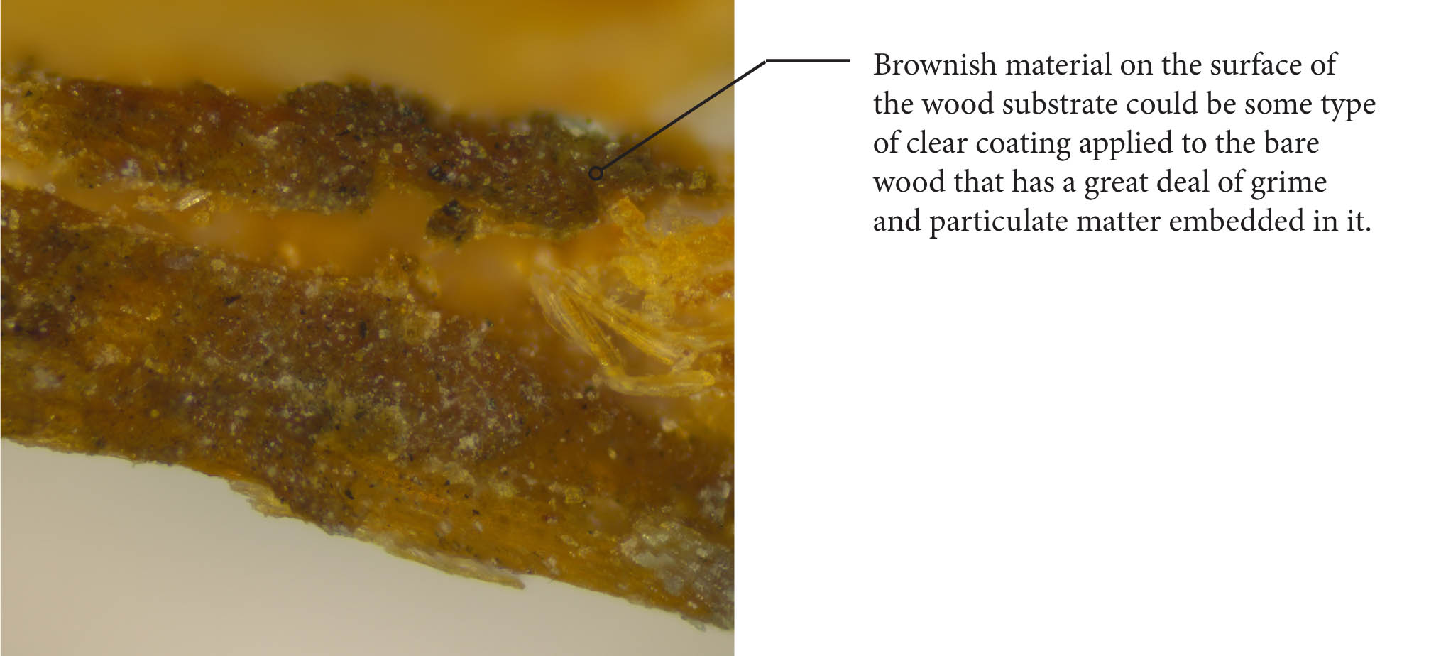 Figure 11. MHS 5, uncast, visible light, 100x
Only early layer on wood shown.
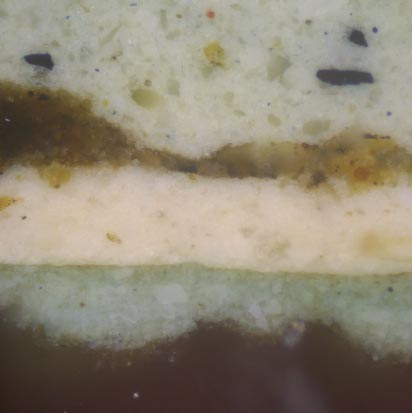
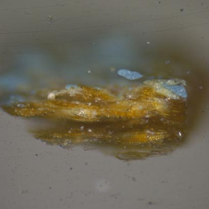
Of all of the generations on the Mayo House stair fragment, the first generation is the most difficult to interpret. While examining the baluster surface, it was observed that the paints have cleaved extensively at the interface between this generation and the second blue paint generation. This suggested that this first generation was not a primer, but an independent finish.
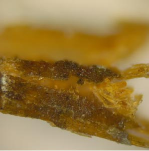
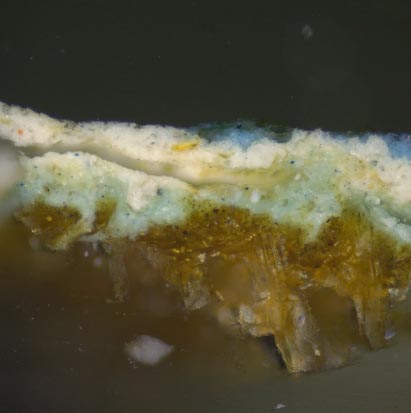
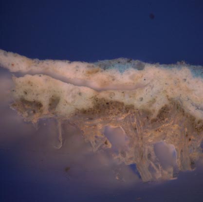
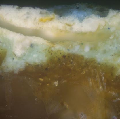
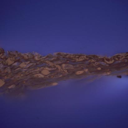
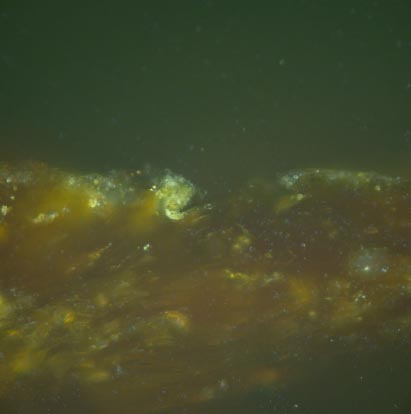
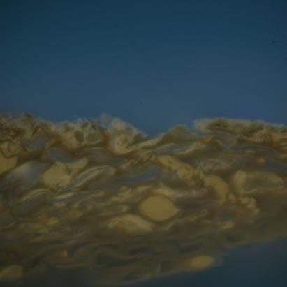
On the uncast sample portions (fig. 11), this generation is a brittle brown material on the surface of the wood. In cross-section (fig. 12), it is a thick, brown-colored layer with black, orange, and colorless particulates embedded throughout. In reflected ultraviolet light, the matrix of this layer has a pale orange autofluorescence, suggesting that a resinous material, possibly shellac, is present. Fluorochrome staining found this generation was positive for carbohydrates, negative for lipids (oils) but a slight positive reaction for proteins was observed (see figs. 15-17). The delivery solvents (acetone for FITC, ethanol for DCF), did appear to partially solubilize this material, which is consistent for shellac coatings or aged resins. Further analysis with an instrumental technique, such as Fourier-transform infrared spectroscopy (FTIR), or gas chromatography — mass spectrometry (GC-MS), is recommended to elucidate this coating.
Taking the data into consideration, it seems possible that this is a resinous material, possibly shellac, that was first applied to the wood surface. It may have been exposed for an extended period of time which allowed a great deal of dirt and dust to embed itself in the coating.
Sample MHS 1: Mayo House stair fragment
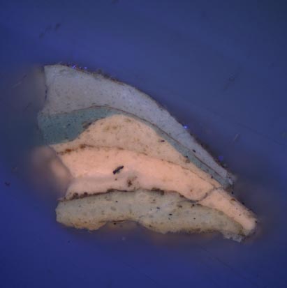
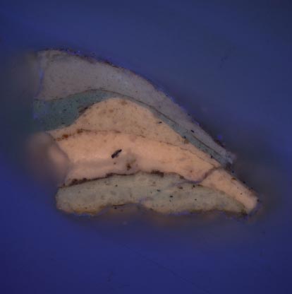
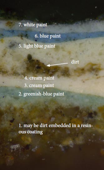 Figure 12a. MHS 1b, visible light, 100x
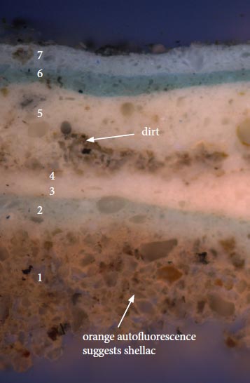 Figure 12b. MHS 1b, UV light, 100x
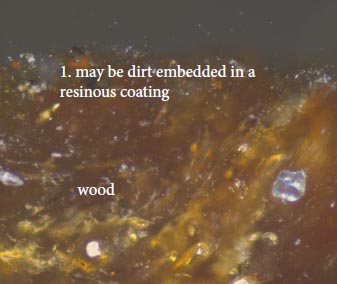 Figure 13a. MHS 1a, visible light, 200x
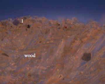 Figure 13b. MHS 1a, UV light, 200x
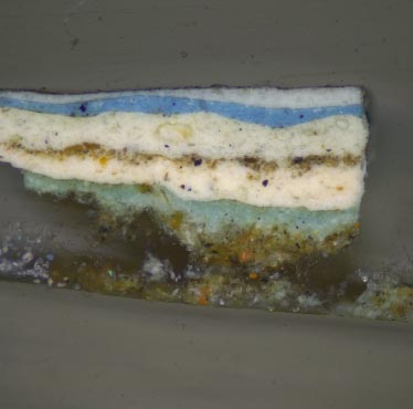
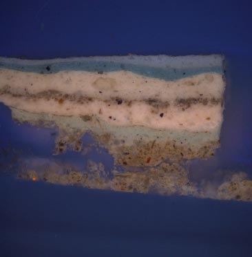
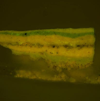
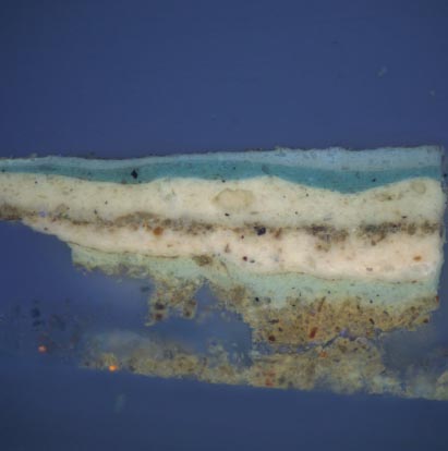
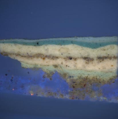
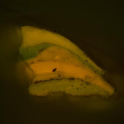
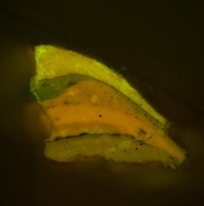
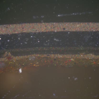
Sample MHS 2: Mayo House stair fragment
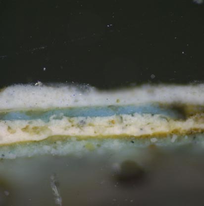
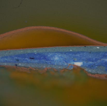
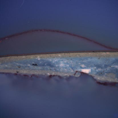
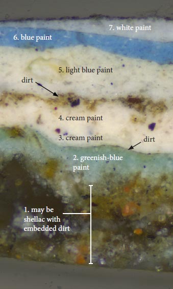 Figure 14a. MHS 2, visible light, 100x
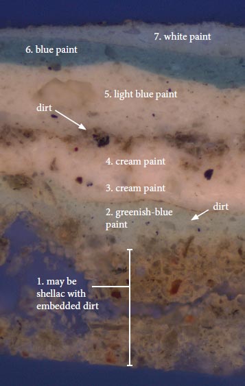 Figure 14b. MHS 2, UV light, 100x
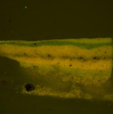
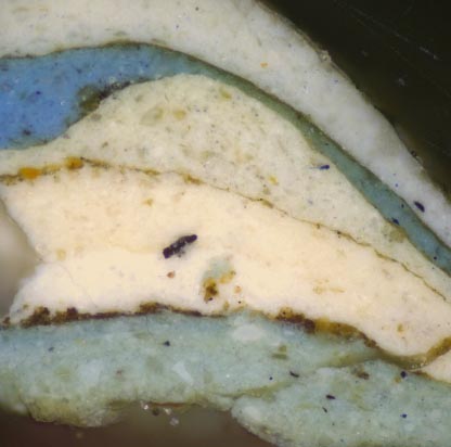
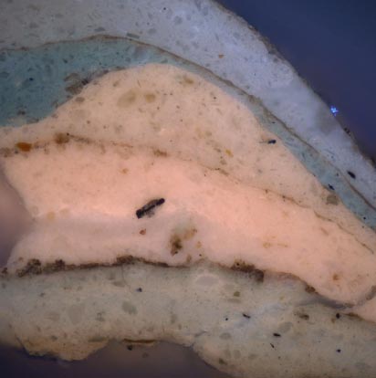
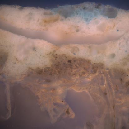
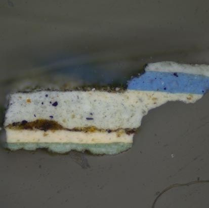
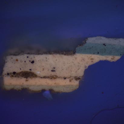
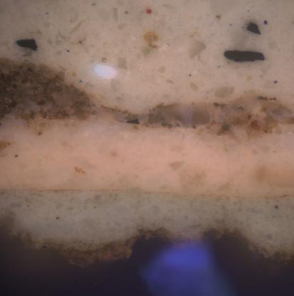
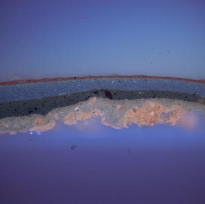
The wood substrate is not shown in this sample, but a thick accumulation of dirt appears to be present on the surface. The matrix of this generation has a pale orange autofluorescence suggesting that a natural resin, such as shellac, is present, identified as generation 1. Examination of an uncast portion of the wood substrate in sample MHS 5 at low power magnification (30x) did reveal a somewhat glossy brown material on the surface of the wood which contained embedded particles of dirt (see fig. 11). The extensive amount of soiling suggests that it was exposed for a long period of time. During this period, the resin coating might have been regularly re-applied, causing the particulate matter to become embedded onto the surface.
Generation 2 is a blue paint covered by a thin layer of grime which indicates an extended period of exposure. Generation 3 and 4 are white paints whose pinkish autofluorescence is suggestive of a lead white in oil, but this would need to be confirmed with an instrumental technique such as SEM-EDS or Raman Spectroscopy. The dirt on the surface of generation 4 suggests it was exposed for a very long period of time. Generation 5 is a light blue paint (which appears white in cross-section). Generation 6 is a deep blue color, and generation 7 is a white paint, which is the current presentation surface.
On the George Pitt House stair, none of these paint generations were found, although an orange autofluorescence was observed in the wood cells, possibly remnants of generation 1 (fig. 23b). The absence of any of the paints seen above suggests that during the restoration, the historic finishes were removed from the rest of the staircase.
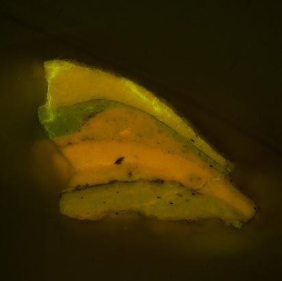
Fluorochrome staining — TTC (carbohydrates)
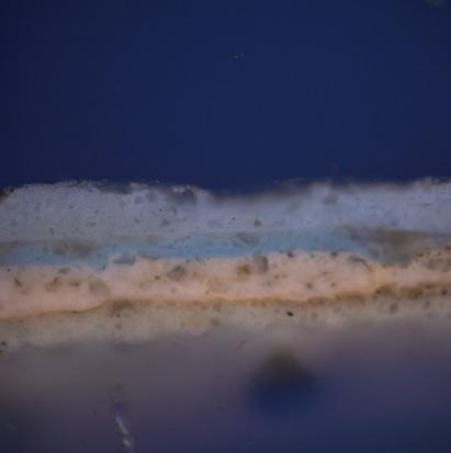
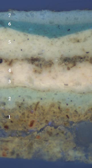 Figure 15a. MHS 2, UV light, 100x. Before TTC for carbohydrates.
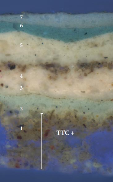 Figure 15b. MHS 2, UV light, 100x. TTC reaction.
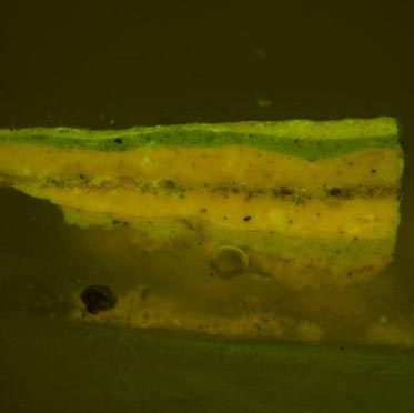
Sample MHS 2 was stained with TTC to tag carbohydrates in the sample. A positive reaction (a reddish-brown color), was observed in generation 1. Since this layer contains soiling materials, this reaction could result from 'starchy' organic matter in this layer. It could also result from a gum additive in the resinous layer applied to the baluster. Generation 1 appears to have been solubilized by the ethanol used to deliver the stain. This is consistent with the suggestion that this layer is shellac-based, or contains an aged natural resin that has become more polar with time.
Instrumental analysis with Fourier-transfrom infrared spectroscopy (FTIR), or gas chromatography-mass spectrometry (GC-MS), would be necessary to more positively identify the organic components of this coating.
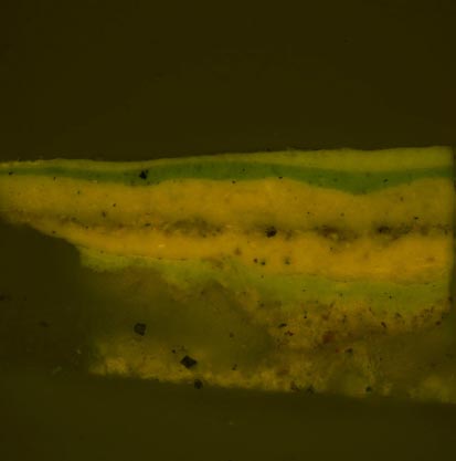
Fluorochrome staining — FITC (proteins)
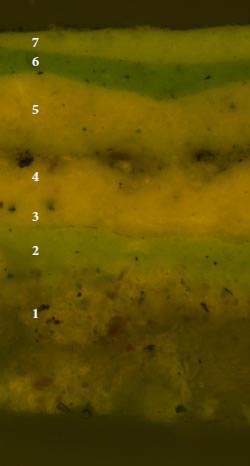 Figure 16a. MHS 2, B2A filter cube,100x. Before FITC stain for proteins.
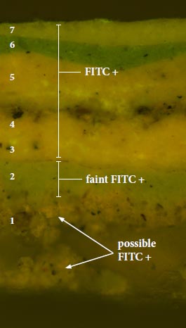 Figure 16b. MHS 2, B2A filter cube,100x. FITC reaction.
Sample MHS 2 was stained with FITC to tag proteins in the sample. It is difficult to interpret the reaction in generation 1, which appears to have been solubilized by the acetone in which the stain was delivered. This is consistent with the suggestion that this layer contains shellac, or an aged natural resin that has increasingly grown more polar with time. Furthermore, polyhydric materials, such as shellacs, can exhibit positive FITC reactions when present in higher concentrations.
Positive reactions (a bright, yellow-green fluorescence) were observed in generations 2-7. These paints also reacted positively for oils with DCF (see next page), suggesting these could be emulsion preparations, containing a mixture of protein and oil. However, this reaction could also be a false positive resulting from urethanes or detergents in modern, industrially-prepared paint formulations. Instrumental analysis with Fourier-transfrom infrared spectroscopy (FTIR), or gas chromatography-mass spectrometry (GC-MS), would be necessary to more positively identify the organic components of these coatings.
Fluorochrome staining — DCF (lipids)
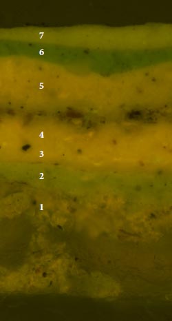 Figure 17a. MHS 2, B2A filter cube,100x. Before DCF
stain for lipids.
Figure 17a. MHS 2, B2A filter cube,100x. Before DCF
stain for lipids.
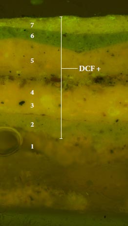 Figure 17b. MHS 2, B2A filter cube,100x. DCF reaction.
Sample MHS 2 was stained with DCF to tag lipids (oils) in the sample. Generation 1 appears to have been solubilized by the ethanol solvent used to deliver the stain. This is consistent with the suggestion that this layer contains shellac, or an aged natural resin that has become more polar with time.
A positive reaction (a bright yellow-green fluorescence), was observed in generations 2-7. These paints also reacted positively for proteins with FITC (see previous page), suggesting these could be emulsion preparations, containing a mixture of protein and oil. This reaction could also be a false positive resulting from detergents or urethane additives in modern, industrially-prepared paint formulations.
Instrumental analysis with Fourier-transfrom infrared spectroscopy (FTIR), or gas chromatography-mass spectrometry (GC-MS), would be necessary to more positively identify the organic components of these paints.
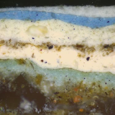
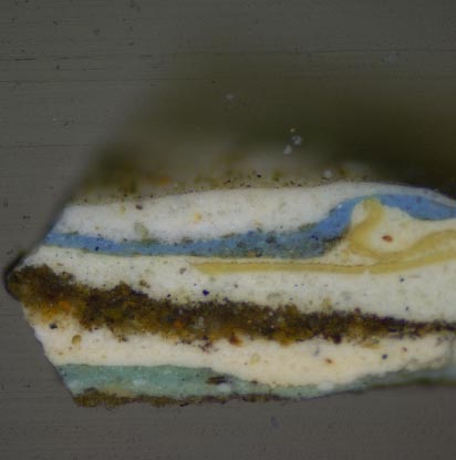
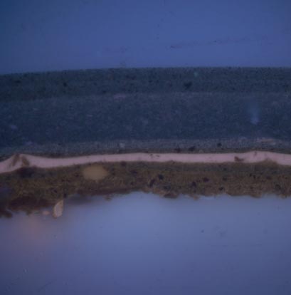
Sample MHS 4: Mayo House stair fragment
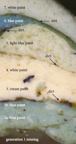 Figure 18a. MHS 4, visible light, 200x
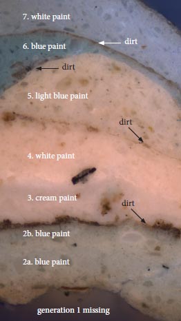 Figure 18b. MHS 4, UV light, 200x
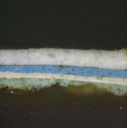
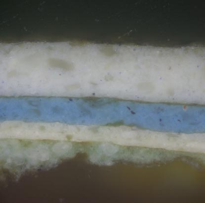
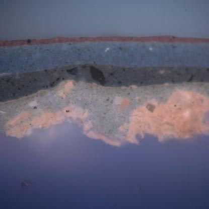
Sample MHS 4 contains the same paint history as the other baluster samples, but more clearly shows that generations two was applied in two coatings, and the hard 'boundary' between generations 3 and 4 indicates they are individual generations.
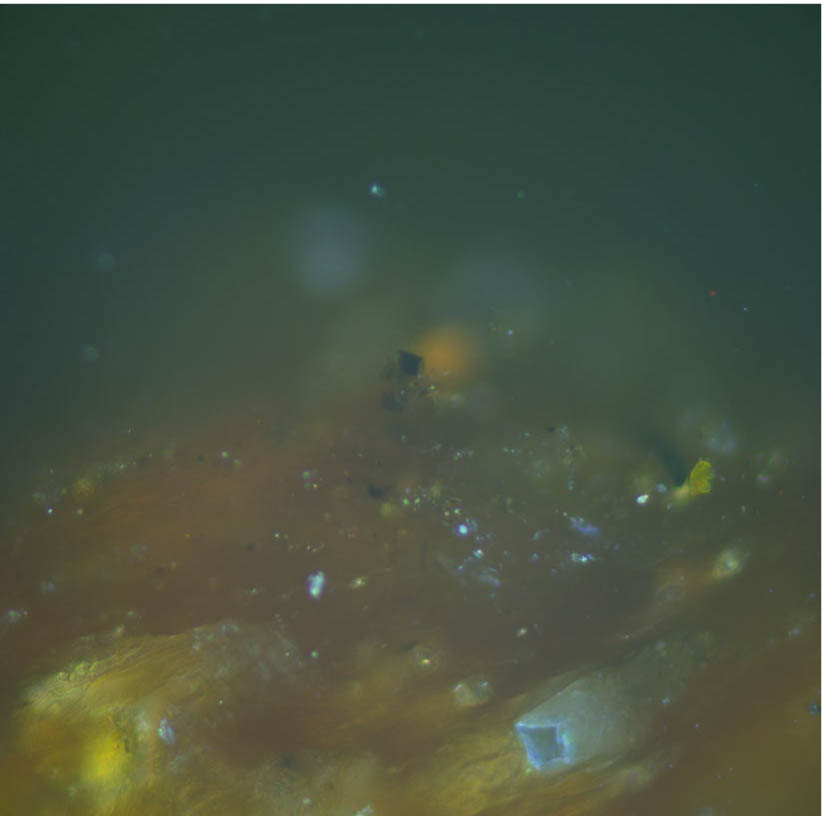
The second generation light blue paint in sample MHS 4 was examined with PLM to determine the pigment composition (see fig. 19).
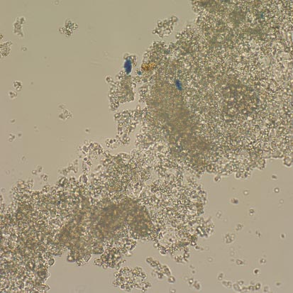
Pigment identification with polarized light microscopy (PLM)
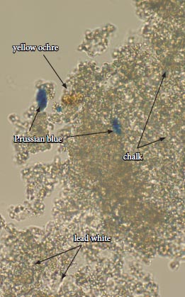 Figure 19a. Dispersed pigment sample from MHS 4, generation 2, light blue paint. Transmitted plane polarized
light, 1000x
Figure 19a. Dispersed pigment sample from MHS 4, generation 2, light blue paint. Transmitted plane polarized
light, 1000x
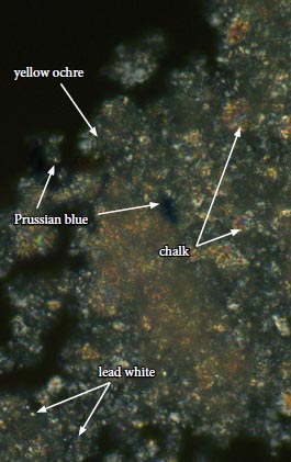 Figure 19b. Dispersed pigment sample from MHS 4, generation 2, light blue paint. Transmitted cross polarized
light, 1000x
Figure 19b. Dispersed pigment sample from MHS 4, generation 2, light blue paint. Transmitted cross polarized
light, 1000x
A sample of the second generation blue paint in sample MHS 4 was collected with a clean scalpel blade, dispersed on a glass slide, and mounted with Cargille Meltmount (refractive index 1.66), for polarized light microscopy.
The majority of pigments are a combination of lead white (2PbCO3 ‧ Pb(OH)2), appearing as small colorless grains that are highly birefringent under crossed polars, and chalk (CaCO3), visible as larger, irregular plates that display a sweeping birefingence under crossed polars.
Small amounts of Prussian blue (Fe4[Fe(CN)6]3) were also present. These particles were observed in a variety of sizes and shapes, which have a deep blue color in transmitted light but are isotropic (dark), under crossed polars. Small amounts of yellow ochre (Fe2O3 ‧ nH2O), appears to have been added to lend the paint a slightly greenish tone. This pigment appears as amorphous agglomerations of particles that are yellow in transmitted plane polarized light, but isotropic (dark), under crossed polars.
Sample PHS 1: George Pitt House stair; half baluster at newel post on landing
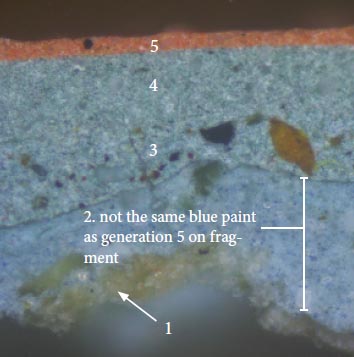 Figure 20a. PHS 1, visible light, 400x
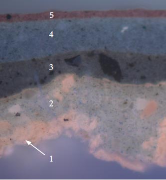 Figure 20b. PHS 1, UV light, 400x
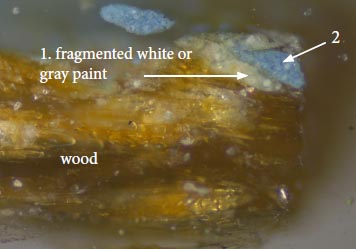 Figure 21a. PHS 1a, visible light, 100x (enlarged)
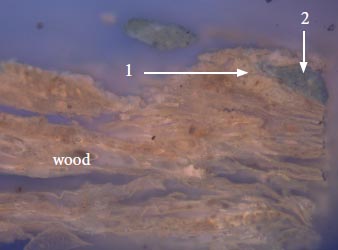 Figure 21b. PHS 1a, UV light, 100x (enlarged)
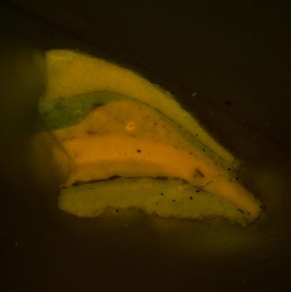
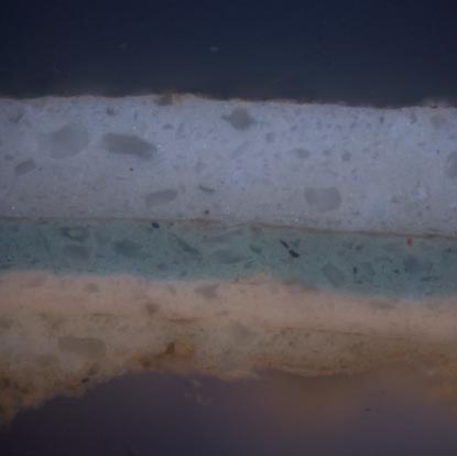
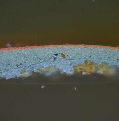
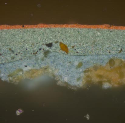
The paint evidence on the George Pitt House stair suggests that five generations are present. The resinous layer on the baluster fragment was not readily apparent here, even when viewed at higher magnifications. The first generation consists of remnants of disrupted, cream-colored or gray layer with a pinkish autofluorescence that might have been applied during the renovation (c.1930), as this layer was not observed on the baluster fragment. It was also seen in sample PHS 3 (fig. 22, 23).
Generation 2 is a bright blue paint that at first glance appears similar to the fifth generation bright blue on the baluster fragment. However, comparison of samples from the George Pitt House stair and the baluster fragment suggest that these are not the same paints. By comparison, the paint on the baluster fragment is a deeper color and much more finely ground. Generations 4-6 have finely-ground pigment particles dispersed within paint films with smooth, even surfaces and a dim autofluorescence suggestive of a synthetic binding medium (late 20th-century or later).
Sample PHS 3: George Pitt House stair; handrail
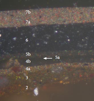 Figure 22a. PHS 3, visible light, 400x
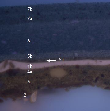 Figure 22b. PHS 3, UV light, 400x
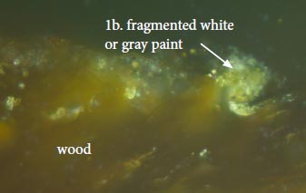 Figure 23a. PHS 3, visible light, 400x
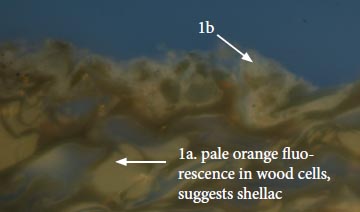 Figure 23b. PHS 3, UV light, 400x
Seven generations of paint were identified on the George Pitt House stair handrail, almost all of which are black or dark-brown paints (the current paint, seen here as generation 7, is dark-brown). The pale orange autofluorescence in the wood cells suggests that a resinous material, possibly shellac, was used to seal the wood before painting (1a). It is unclear if this is the same resinous sealant found on the Mayo House baluster fragment. This is followed by remnants of the cream or gray paint (1b), that was also found in sample PHS 1 (fig.21). With the exception of generation 4b, all are smooth, consistent paint films with finely ground pigment particles well-dispersed throughout their matrices. Their dim autofluorescence is suggestive of modern (twentieth-century), synthetic binding media, particularly in generations 5-7. The brighter autofluorescence of generation 4b suggests that it is a varnish layer that was applied over generation 4a, a red-brown paint, to give it a protective, glossy surface.
In consideration of this evidence it appears that the earliest finishes were removed when the staircase was reinstalled in the reconstructed George Pitt House.
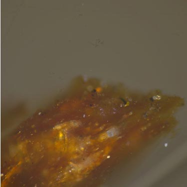
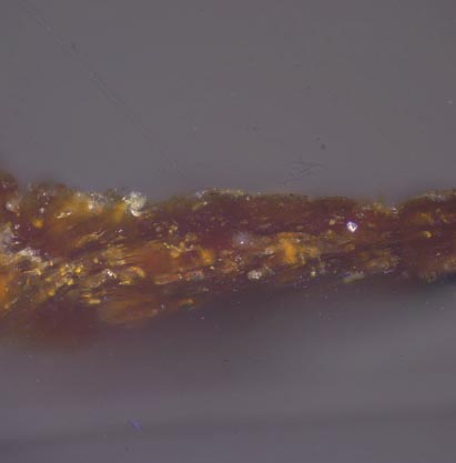
Mayo House stair fragment: Colorimetry
Generation 2: light blue paint
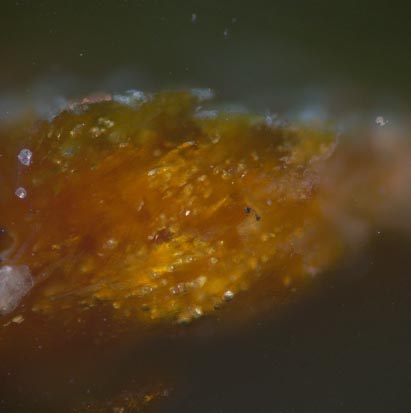
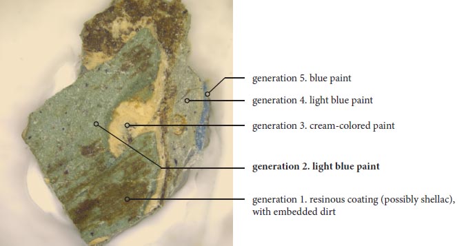 Figure 24a. MHS 8, uncast, visible light, 40x
Accurate color readings for the second generation blue paint could not be obtained with the Minolta Chroma Meter colorimeter/microscope. Instead, the closest commercial color match was determined by eye using a stereomicroscope at 30x magnification with a color corrected light source. The closest commercial color match was determined to be Colonial Williamsburg swatch CW 501 "Wythe House Blue."
 Colonial Williamsburg Paint
CW-501
"Wythe House Blue"
| CIE L*a*b* values | L* | a* | b* |
|---|---|---|---|
| 63.58 | -9.09 | -6.03 | |
| Munsell values | hue | value | chroma |
| 2.3B | 6.3 | 2.4 |
Conclusions:
This study found seven paint generations on the baluster fragment from the Mayo House stair, which predate the restoration period (1930s). None of these paints were found on the rest of the staircase that is currently installed in the George Pitt House. Instead, up to seven generations of mostly modern, industrially-prepared paints and varnish were found. This comparison suggests that the staircase was stripped of its original finishes when it was transferred to its new location in 1936. Samples from both the Mayo House baluster fragment and the half-baluster on the George Pitt House staircase did exhibit an orange autofluorescence in the wood fibers, possibly a shellac coating that was the first period finish. However, it could not be conclusively determined that the shellac on the George Pitt House stair is the same as the shellac on the baluster fragment.
The first period coating on the Mayo House baluster fragment may be a resinous coating, possibly shellac, that would have protected the wood but left the natural grain exposed as a presentation surface. However, further analysis with FTIR or GC-MS is recommended to identify this material with confidence. There is a large amount of embedded particulate matter in this coating, suggesting it was exposed for a long period of time. The second generation is a greenish-blue paint made with lead white, chalk, Prussian blue, and yellow ochre. All of these pigments were readily available in eighteenth-century Williamsburg. Fluorochrome staining suggests the binding media may contain both lipidic (oil) and proteinaceous components. A color match for this paint is provided on page 18. It is unclear precisely when this greenish-blue paint was applied, but the significant amount of dirt on the wood substrate suggests it was well after the staircase had been completed.
Generations 3 and 4 are cream-colored paints and generation 5-7 are light blue, deep blue, and white paints, respectively. All stained positively for lipidic (oil) and proteinaceous components. It is unclear if the binding media of these paints are emulsions or if these reactions are 'false positives' generated by additives in industrially-prepared paint formulations.
It is important to remember these paints are specific to the balusters only, and do not necessarily reflect the paint history of the staircase overall. In fact, an archival photograph of the Mayo House interior prior to demolition shows the stair balusters painted a light color with darker newel posts and handrail (see page 4). This polychrome treatment might have been carried over from the 18th century, but without additional physical evidence the overall paint scheme cannot be determined.
The finish evidence on the George Pitt House staircase was very different from the baluster fragment. The sample from the half-baluster contained five paint generations in total, and the handrail contained seven generations. On the handrail, the remnants of shellac were found deep in the wood cells, but it is unclear if this is a surviving 18th-century coating or one that was applied much later. The remaining paints appear to date from the 20th century. This comparative study suggests that the Mayo House staircase was stripped of its original finishes when it was moved to the George Pitt House.
References
- Kocher and Dearstyne. 1954. George Pitt house (LT) architectural report, block 18, building 4B, lot 47. (Originally entitled "Architectural report Pitt-Dixon house block 18, colonial lot #47"). Unpublished report for the Colonial Williamsburg Foundation, Architectural Research Department.
- Eastaugh, N., et. al. 2008. Pigment Compendium: a dictionary and optical microscopy of historical pigments. Oxford, Butterworth-Heinemann.
- Gettens, R., and G. Stout. 1942. Painting materials: a short encyclopedia. New York, Dover Publications, Inc.
- Whiffen, M. 1984, revised ed. The eighteenth-century houses of Williamsburg. Virginia. The Colonial Williamsburg Foundation: 228-233.
- Wolbers, R. 2008. Color and light. Lecture for General Conservation Science ARTC 615, at the Winterthur/ University of Delaware Program in Art Conservation.
Appendix A. Sampling and Analytical Procedures
Sample Preparation:
The samples were cast in mini-cubes of Extec Polyester Clear Resin (methyl methacrylate monomer), polymerized with the recommended amount of methyl ethyl ketone peroxide catalyst. The resin was allowed to cure for 24 hours under ambient light. After cure, the individual cubes were removed from the casting tray and sanded down using a rotary sander with grits ranging from 200 - 600 to expose the cross-section surface. The samples were then dry polished with silica-embedded Micro-mesh Inc. cloths with grits ranging from 1500 to 12,000, lending the final cross-section surface a glassy-smooth finish.
Microscopy and Documentation:
The cross-section samples were examined using a Nikon Eclipse 80i microscope equipped with an EXFO X-cite 120 fluorescence illumination system fiberoptic halogen light source. Samples were examined and photographed under visible and ultraviolet light conditions (330-380 nm), at 20 to 200x magnifications. Digital images were captured using a Spot Flex digital camera with Spot Advance (version 4.6) software. All images were recorded as 12.6 MB tiff files and stored on a hard drive in a folder titled "Finnie Interior" on Susan Buck's laboratory computer. A separate set of images will be stored on the CWF digital database, accompanied by a digital version of the final report.
Information Provided by Visible and Ultraviolet Light Microscopy:
When examining paint cross-sections under reflected visible and ultraviolet light conditions, a number of physical characteristics can be observed to assist with the interpretation of a paint stratigraphy. These include the number and color of layers applied to a substrate, the thickness or surface texture of layers, and pigment particle size and distribution within the paint film. Relative time periods for coatings can sometimes be assigned at this stage: for instance, pre-industrial-era paints were hand ground, lending them a coarse, uneven surface texture with large pigment particles that vary in size and shape. By contrast, more "modern", industrially-prepared paints have smoother, even surfaces and machine-ground pigment particles of a consistent size and shape. Furthermore, he presence of cracks, dirt layers, or biological growth between layers can indicate presentation surfaces and/or coatings that were left exposed for an extended period of time.
Under UV light conditions, the presence and type of autofluorescence colors can distinguish sealants, clear coatings, and binding media, from darker dirt or paint layers within the stratigraphy. For instance, shellacs exhibit a distinct orange-colored autofluorescence, while natural resins (such as dammar and mastic), typically fluoresce a bright white color. Oil media tends to quench autofluorescence, while most modern, synthetic paint formulations (such as latex) exhibit no fluorescence at all. Some pigments, such as verdigris, madder, and zinc white, have distinct fluorescence characteristics, as well. UV light microscopy is critical to help distinguish otherwise identical layers often found in architectural samples- such as successive varnishes, or multiple layers of unpigmented (white) limewash.
Binding Media Analysis using Fluorochrome staining:
Fluorochrome stains adapted from the biological sciences were used to characterize the paint binding media (oils, proteins, carbohydrates), in layers within the cross-section sample. The following stains were used in this analysis:
2,7 Dichlorofluorescein (DCF): 0.02% w/v in ethanol. Fluorescent labeling reagent for lipids, particularly drying oils. One drop of stain was applied to the surface of the sample, blotted immediately, and cover-slipped with mineral spirits. The reaction was observed using the B-2A filter cube (450-490 nm). A positive reaction is a bright yellow-green fluorescence.
Triphenyl tetrazolium chloride (TTC): 1.0% w/v in ethanol. Labeling reagent for carbohydrates (gums, starches, cellulosic thickeners). One drop of stain was applied to the surface of the sample, blotted dry, and allowed to sit for approximately 45 seconds before cover-slipping, (must be allowed to react with atmospheric moisture for reaction to move forward). The reaction is observed under reflected UV light conditions (330-380 nm). A dark red-brown color is seen where carbohydrates are present.
Fluorescein isothiocyanate (FITC): 0.02% w/v in anhydrous acetone. Fluorescent labeling reagent for proteins. One drop of stain was applied to the surface of the sample, blotted immediately, and cover-slipped with mineral spirits. The reaction was observed using the B-2A filter cube (450-490 nm). A positive reaction is a bright yellow-green fluorescence.
Pigment Identification with Polarized Light Microscopy:
To collect a pigment sample for polarized light microscopy (PLM), a surgical scalpel was used to collect a small scraping from a clean, representative area of paint. The blade was then pressed and pulled across a clean glass microscope slide, dispersing the pigment particles across the surface. The pigments were then permanently embedded under a cover slip using Cargille Meltmount (refractive index 1.66). The embedded pigments were then examined in cross and plane-polarized transmitted light with the Nikon Eclipse 80i microscope at 1000x magnification (using an oil immersion objective). The observed morphologies (size, shape, agglomeration, cleavage patterns), and optical properties (including color, refractive index, extinction), were compared to reference standards as well as literature sources before making final determinations.
Color measurement and matching:
Color measurements were taken using the Minolta Chroma Meter CR-241 colorimeter/microscope in Susan Buck's paint analysis laboratory. Equipped with an internal 360-degree pulsed xenon arc lamp, this instrument is capable of obtaining accurate color measurements in any one of five different tristimulus color measurement systems from areas as small as 0.3mm. For the purposes of this project, color values in CIE L*a*b* colorspace and the Munsell color system were obtained.
The CIE L*a*b* color space system (developed in 1976 by the Commission International de l'Eclairage, and now an internationally accepted industry-standard color measuring system) uses three numerical values, known as "tristimulus" values, to measure color: L* is the lightness variable, representing dark to light on a scale of 0-100, while a* and b* are chromaticity coordinates, a* representing red to green on scale from -50 to +50, and b*representing blue to yellow on a scale from -50 to +50. These three coordinates are used to plot the location of a color in the CIE L*a*b* colorspace.
23These resulting values can be used to quantify color differences (ΔE), between two samples. To obtain this value, the following calculation is used: ΔE = (ΔL*)½ + (Δa*)½ + (Δb*)½
Generally, a ΔE value ≤ 3 cannot be perceived by the human eye (Wolbers 2008). Therefore, for any two samples, ΔE values at or below this range are considered acceptable matches.
Ideally, color measurements should be collected from a clean, unweathered sample area. If necessary, a scalpel is used to scrape an area clean before color matching. Due to inherent color variations in paints (especially in hand ground, pre-industrial coatings), multiple readings are taken and averaged together to establish the final CIE L*a*b* values.
Due to the deterioration, soiling, and fragmentary nature of some early paints, color readings cannot always be obtained with the Chroma Meter. In these instances, paints are matched by eye to Munsell standard color swatches and commercial paint chips, using a stereomicroscope at 30x magnification with a color corrected light source. Commercial systems consulted include Benjamin Moore, Sherwin Williams, Pittsburgh Paints, and the Colonial Williamsburg Color Collection.
Appendix B. Sampling Memorandum
| Date: | Monday, September 27, 2010 |
|---|---|
| Name: | Kirsten Travers |
| Site: | George Pitt House, Block 18 Building 4 |
On the above date the George Pitt house was visited by Kirsten Travers, Matt Webster, and Dani Jaworski. The house is a reconstruction but contains an 18th century stairwell from another building, the Mayo House (now destroyed).
The purpose of the visit to the reconstructed house was to collect samples to search for intact paint sequences. The stair is currently painted a dark red with a glossy, dark brown hand rail. No paint buildup could be seen with the naked eye. The current resident of the house said that the stair was previously painted a bright blue. In-situ, small excavations were made with a microscalpel, and examination at 30x magnification suggested that much of the early paint history was removed, as only the modern red and blue layers were visible.
A baluster from the original stair (now housed in the CWF architectural fragments collection, May-01-08, AF-CHES 90.0226, EEK, 1-17-08), was brought to the site by Dani Jaworski for comparison. The current presentation surface is a soiled, off-white color, but there are visible accumulations of thick, flaking paint layers revealing white and blue paints underneath. With the naked eye, one can see a thin, reddish-brown coating applied directly to the wood substrate. This fragment was made available to Kirsten Travers to sample and may generate intact paint sequences. The results may inform the decorative history of the Mayo House stair, and may offer insight into the treatment of the relocated stair.
‧ Three (3) samples were taken from the in-situ Pitt House stair:
PHS_1: Stair landing, half baluster against the landing newel post (going UP). Bottom third section of the baluster, Top cap of the urn (just below the square base of the mid-section).
—Current presentation surface is red. I did get some wood substrate with associated layers. All I saw in-situ was modern paint (blue, followed by red), applied directly to the wood. Some whitish, coarse material is visible on some of the wood fibers (could be a putty).
PHS_2: Second flight, top molding of stringcourse, just below 6th baluster.
—Current presentation surface is red. Sample site already had large crack/loss that was painted over. Dark reddish-brown paint applied directly to the wood, similar to that seen on the fragment, was observed here. Besides that, only modern layers (the blue and red) were observed.
PHS_3: Second flight, top rail. Flat face just below the cap and up against the newel post. Side facing the wall.
—Current presentation surface is dark, glossy brown. Removed wood substrate and paint layers, but layers cleaved after sampling, so sample will have to be "mended" in photoshop. All I saw were modern, dark brown, gray, and black paints.
25‧ Eight (8) samples were taken from the Mayo house stair fragment, [AF-CHES 90.0226]. Almost all of the samples cleaved at the interface between paint and wood. In every instance I tried to get a good sample of the wood fibers as well.
MHS_1: Top surface of baluster base, 6 ½" up from bottom of the piece (bottom is measured from the longest edge of the base).
MHS_2: Top surface of the "urn" shaped element, 8" up from bottom of piece.
MHS_3: Top surface of base (the part that is supporting the "column-like element" on the baluster), 12" up from bottom.
MHS_4: Capital of long shaft-like element, 21" from bottom.
MHS_5: Shaft, 19" from bottom.
MHS_6: Body of "urn" shaped element, 8 ¾" from bottom. This sample is very fragmented.
MHS_7: Face of the pedestal base. 5 ¾" up from bottom.
MHS_8: Face of pedestal base, very top near unfinished edge. 4 ½" away from sample MHS_6.
 MHS 5 uncast early layer on wood 100x
 MHS 8 uncast 40x
 MHS 1 VIS 100x
 MHS 1 UV 100x
 MHS 1 VIS 200x
 MHS 1 UV 200x
 MHS 1a VIS 200x
 MHS 1a UV 200x
 MHS 2a VIS 200x
 MHS 2a UV 200x
 MHS 2a VIS 400x
 MHS 2a UV 400x
 MHS 2 VIS 100x
 MHS 2 UV 100x
 MHS 2 VIS 200x
 MHS 2 UV 200x
 MHS 2 B2A b4DCF 100x
 MHS 2 B2A DCF 100x
 MHS 2 B2A b4FITC 100x
 MHS 2 B2A FITC 100x
 MHS 2 UV b4TTC 100x
 MHS 2 UV TTC 100x
 MHS 3 VIS 100x
 MHS 3 UV 100x
 MHS 3 VIS 200x
 MHS 3 UV 200x
 MHS 4 B2A b4DCF 100x
 MHS 4 B2A b4FITC 100x
 MHS 4 B2A DCF 100x
 MHS 4 B2A FITC 100x
 MHS 4 gen 1 blue BF 1000x
 MHS 4 gen 1 blue DF 1000x
 MHS 4 VIS 100x
 MHS 4 UV 100x
 MHS 4 VIS 200x
 MHS 4 UV 200x
 MHS 4 UV b4TTC 100x
 MHS 4 UV TTC 100x
 MHS 5 VIS 200x
 MHS 5 UV 200x
 MHS 5 VIS 400x
 MHS 5 UV 400x
 MHS 6 VIS 200x
 MHS 6 UV 200x
 MHS 6 VIS 400x
 MHS 6 UV 400x
 MHS 7 VIS 200x
 MHS 7 UV 200x
 MHS 7 VIS 400x
 MHS 7 UV 400x
 MHS 8 VIS 100x
 MHS 8 UV 100x
 MHS 8 VIS 400x
 MHS 8 UV 400x
 MHW 7 VIS 400x
 PHS 1 VIS 200x
 PHS 1 UV 200x
 PHS 1 VIS 400x detail of blue
 PHS 1 UV 400x
 PHS 1a VIS 100x
 PHS 1a UV 100x
 PHS 2 VIS 100x
 PHS 2 UV 100x
 PHS 2a VIS 200x
 PHS 2a UV 200x
 PHS 3 VIS 400x
 PHS 3 UV 400x
 PHS 3a VIS 200x
 PHS 3a UV 200x
 PHS 3a VIS 400x
 PHS 3a UV 400x